- Visibility 16 Views
- Downloads 3 Downloads
- DOI 10.18231/j.ijohd.2021.043
-
CrossMark

Early intervention of anterior cross bite malocclusion relating to functional class iii malocclusion
Introduction
The origin of anterior crossbites could be either dental or skeletal. The etiology of anterior dental crossbites is due to the abnormal axial inclination of the maxillary anterior teeth.
Anterior skeletal crossbites are associated with a skeletal problem, such as mandibular prognathism and midface deficiency.[1]
The incidence of anterior dental cross bite is 4-5% and is usually as a result of ectopic eruption or palatal malposition of the maxillary incisors[2] resulting from a lingual eruption path. Other etiological factors include trauma to the primary maxillary incisors resulting in lingual displacement of the permanent tooth buds; presence of supernumerary anterior teeth; crowding in the incisor region, an over-retained, necrotic or pulpless deciduous tooth or root; delayed exfoliation of the primary incisors; and odontomas.[3], [2], [4], [5]
Case Presentation
A 13-year-old boy was referred to the orthodontic clinic with the chief complaint of irregularly placed upper front teeth and an unaesthetic appearance of the maxillary central incisors that were behind the lower anterior teeth. No relevant medical and dental history, and the patient did not have a family history of Class-III malocclusion.
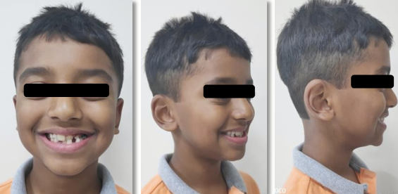
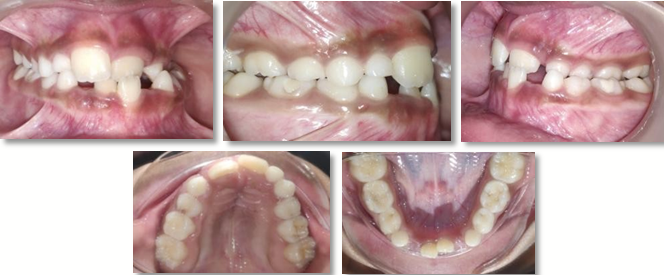
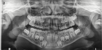
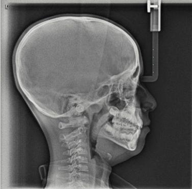
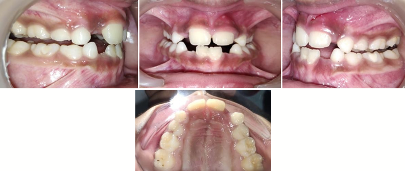
Pre-treatment extra-oral on smiling ([Figure 1]) shows unilateral crossbite of 21 with respect to 31, constricted maxillary arch. On intra-oral examination ([Figure 2]) the permanent maxillary left central incisors were in crossbite, and constricted maxillary arch. The patient was in early-mixed dentition and had a Class-I molar relationship on both sides, with a 2 mm overjet and 80% overbite. The maxillary dental midline was coincident with the facial midline; however, the mandibular dental midline deviated approximately 4 mm to the left. A panoramic radiograph showed early mixed dentition([Figure 3]) and lateral cephalometric radiographic view showed no evidence of basal problem between mandibular and maxillary arches ([Figure 4]).
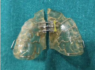
The treatment objectives includes correction of the anterior crossbite, to achieve normal overbite and overjet, alignment of anterior teeth and correction of unilateral crossbite and to improve the patient's facial and dental esthetics. For alignment and correction of the crossbite, a removable acrylic appliance with expansion screw with a posterior bite-opening platform was used.
A screw incorporated in the appliance platform was activated 0.25 mm every 4 days for 16 weeks.([Figure 5]) After 2 months, the maxillary and mandibular incisors
displayed an edge-to-edge bite relationship, and the crossbite was corrected in next 2 months ([Figure 6]) The posterior bite-opening platform was then removed, and screw activation continued every 7 days for another 2 months in order to establish a normal overjet. After 8 months of active treatment, the crossbite of all maxillary incisors and unilateral crossbite was corrected.([Figure 7])

Discussion
Various techniques used to correct anterior dental crossbite are tongue blades, composite inclined planes, reversed stainless steel crowns, removable acrylic appliances with lingual springs and fixed appliances.[3], [2], [4], [5], [6], [7], [8] Factors that are taken into consideration along with the age of the child, are the number of teeth requiring repositioning, overbite, the total number of teeth involved and how parents or child was motivated.[7], [8], [9]
Conclusion
An anterior crossbite affecting two or more teeth or presenting with a reverse overjet in the absence of a functional displacement, may signify an underlying skeletal discrepancy. An anterior crossbite affecting two or more teeth or presenting with a reverse overjet in the absence of a functional displacement, may signify an underlying skeletal discrepancy. An anterior crossbite affecting two or more teeth or presenting with a reverse overjet in the absence of a functional displacement, may signify an underlying skeletal discrepancy.
The timing of orthodontic interventions is important in success of treatment i.e. when to plan the early treatment or indeed to stop skeletal discrepancies altogether three spatial planes. Correct intervention timing will certainly reduce the severity of malocclusion.
Source of Funding
None.
Conflict of Interest
None.
References
- R E Moyers. Handbook of Orthodontics. 1973. [Google Scholar]
- P W Major, K Glover. Treatment of anterior cross-bites in the early mixed dentition. J Can Dent Assoc 1992. [Google Scholar]
- J H Park, T W Kim. Anterior crossbite correction with a series of clear removable appliances: A case report. J Esthet Restor Dent 2009. [Google Scholar] [Crossref]
- S Bayrak, E S Tunc. Treatment of anterior dental crossbite using bonded resin- composite slopes: Case reports. Eur J Dent 2008. [Google Scholar]
- K Heikinheimo, K Salmi, S Myllärniemi. Long term evaluation of orthodontic diagnoses made at the ages of 7 and 10 years. Eur J Orthod 1987. [Google Scholar] [Crossref]
- G Vadiakas, A D Viazis. Anterior crossbite correction in the early deciduous dentition. Am J Orthod Dentofacial Orthop 1992. [Google Scholar] [Crossref]
- H A Kiyak. Patients’ and parents’ expectations from early treatment. Am J Orthod Dentofacial Orthop 2006. [Google Scholar] [Crossref]
- S Sari, H Gokalp, S Aras. Correction of anterior dental crossbite with composite as an inclined plane. Int J Paediatr Dent 2001. [Google Scholar] [Crossref]
- T P Croll, R E Riesenberger. Anterior crossbite correction in the primary dentition using fixed inclined planes. II. Further examples and discussion. Quintessence Int 1988. [Google Scholar]